 Barnorama Welcome To The Fun Barn
The 2025 Nikon Small World Awards celebrate photographers who blend science with artistry in ways that mesmerize and educate.
3rd Place: John-Oliver Dum, Medienbunker Produktion, Bendorf, Rheinland Pfalz, Germany
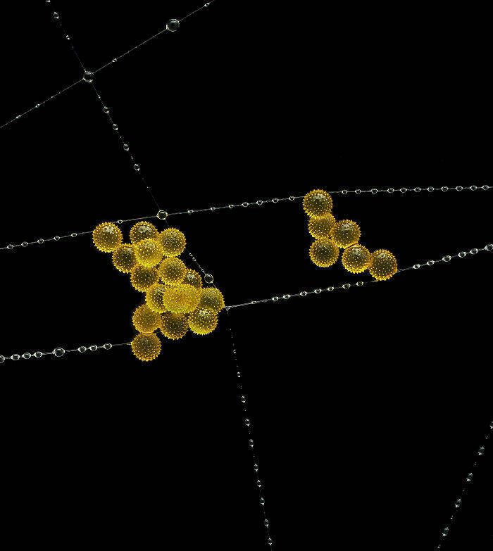
“Pollen in a garden spider web.”
Image Of Distinction: Dr. David Maitland, Art Of Science, St. Andrews, Fife, United Kingdom
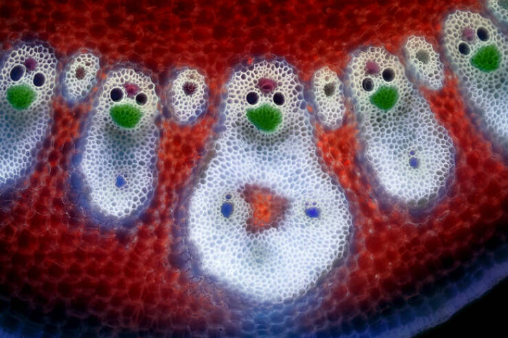
“Vascular bundles in a bamboo leaf (Phyllostachys sp.).”
Image Of Distinction: Ye Fei Zhang, Jiang Yin, Jiang Su, China

“Butterfly (Artopoetes pryeri) eggs.”
Image Of Distinction: Marek Miś, Marek Miś Photography, Suwalki, Podlaskie, Poland
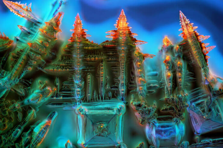
“Crystallized soy sauce.”
Honorable Mention: Gregory B. Murray, Pritchard, British Columbia, Canada
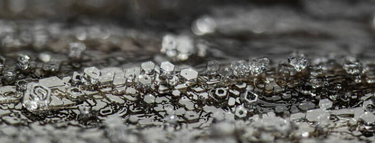
“Frost on a wooden railing.”
2nd Place: Dr. Jan Rosenboom, Rostock, Mecklenburg Vorpommern, Germany
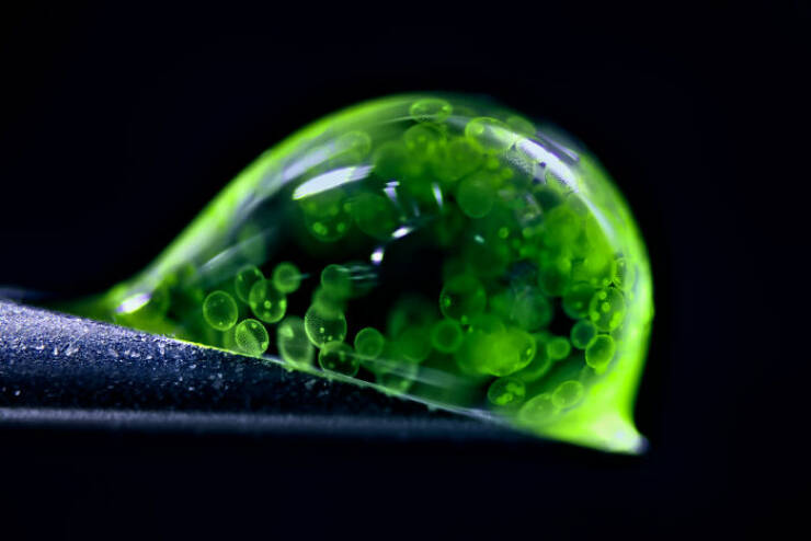
“Colonial algae (Volvox) spheres in a drop of water.”
1st Place: Zhang You, Kunming, Yunnan, China
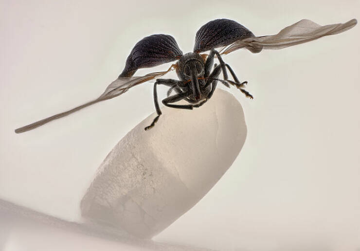
“Rice weevil (Sitophilus oryzae) on a grain of rice.”
5th Place: Dr. Igor Siwanowicz, Howard Hughes Medical Institute (Hhmi), Janelia Research Campus, Ashburn, Virginia, USA

“Spores (blue/purple structures) of a small tropical fern (Ceratopteris richardii).”
11th Place: Marek Miś, Marek Miś Photography, Suwalki, Podlaskie, Poland
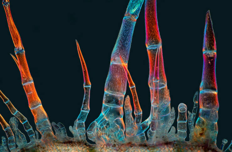
“Sunflower trichomes (hair-like plant outgrowths).”
Honorable Mention: Michael Robert Peres, Rochester Institute Of Technology, School Of Photographic Arts And Sciences, Rochester, New York, USA
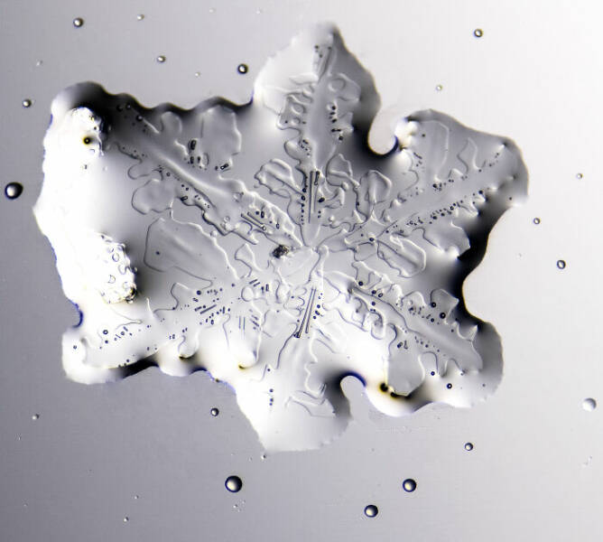
“Melting snowflake.”
14th Place: Manfred Heising, Lwl Museum Of Natural History Münster, Münster, Northrhine-Westphalia, Germany
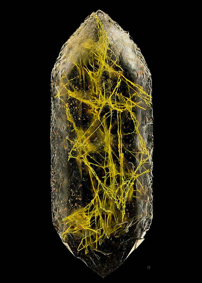
“Quartz with biotic goethite filaments.”
Honorable Mention: Michael Parra Puentes, Bogotá, Cundinamarca, Colombia
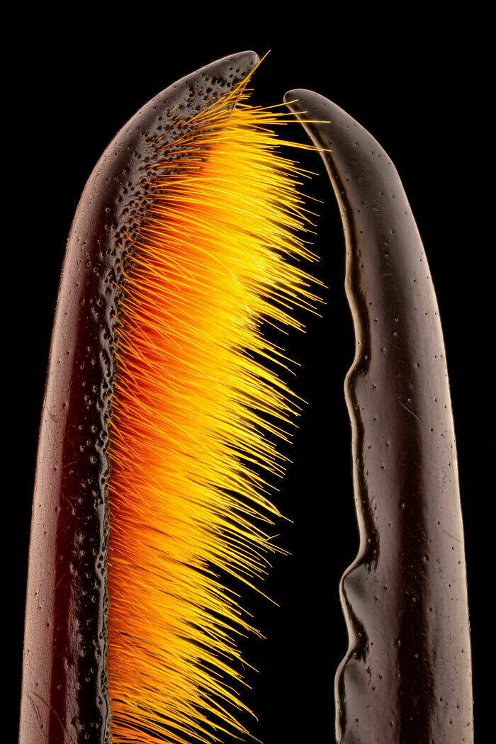
“Thoracic and cephalic horn of a male beetle (Golofa porteri).”
Image Of Distinction: Karl Deckart, Eckental, Bavaria, Germany
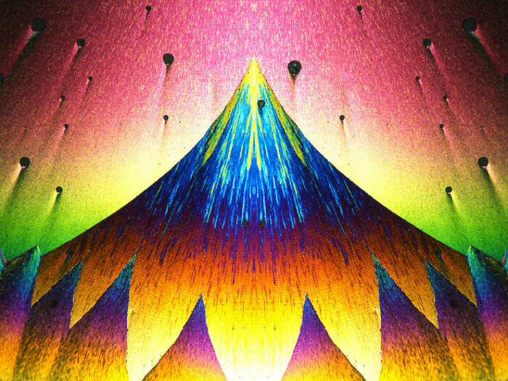
“Recrystallization of phenyl imidazol.”
13th Place: Henri Koskinen, Helsinki University, Helsinki, Uudenmaan Lääni, Finland
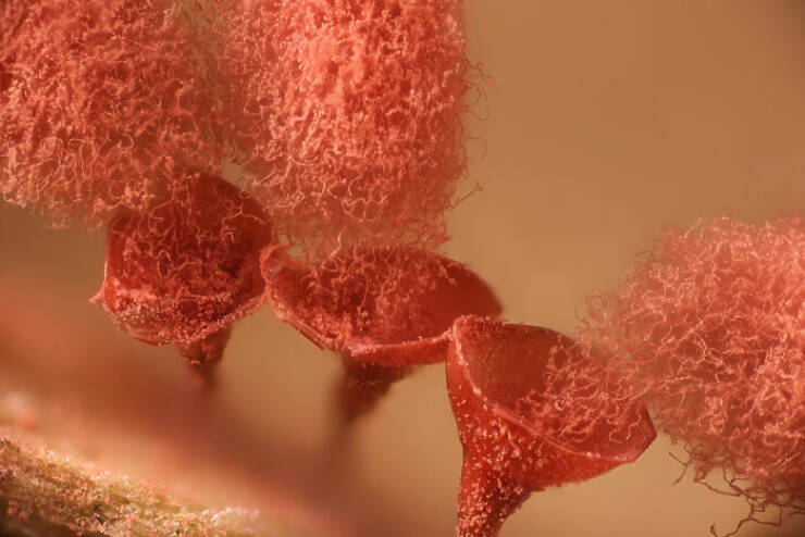
“Slime mold (Arcyria major) releasing spores.”
Honorable Mention: Jiri Cerny, Institute Of Molecular Genetics Of The Czech Academy Of Sciences, Light Microscopy Core Facility, Prague, Czech Republic
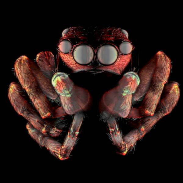
“Jumping spider.”
Image Of Distinction: Daniel Evrard, Aywaille, Liege, Belgium
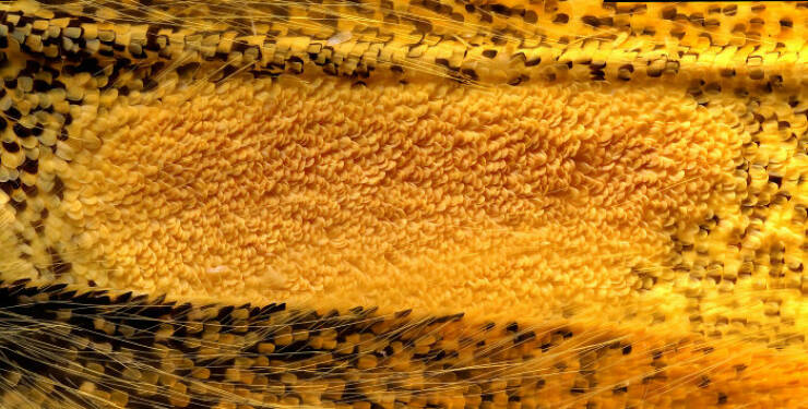
“Androconial (pheromone producing) area of a butterfly (Colias) wing.”
Image Of Distinction: Dr. Francisco Lázaro-Diéguez, Albert Einstein College Of Medicine, Bronx, New York, USA
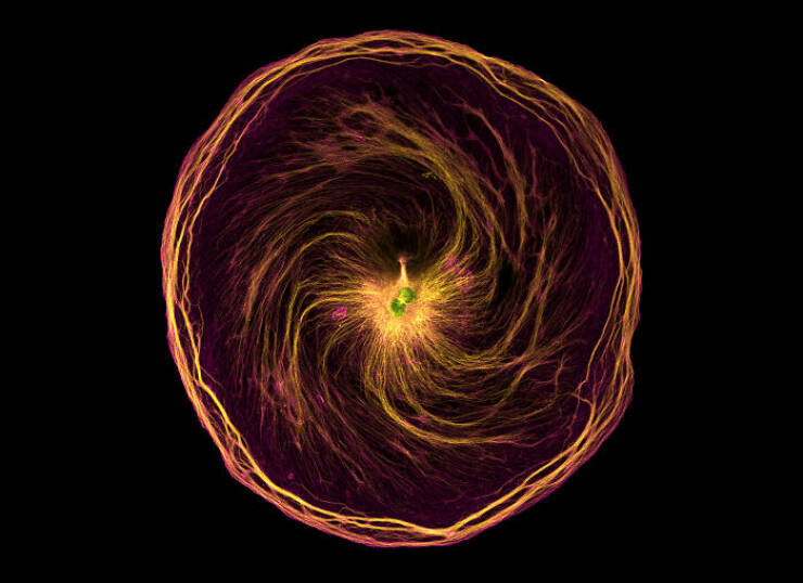
“Dedifferentiated liver cell.”
Image Of Distinction: Dr. Gonzalo Quiroga Artigas, Crbm-Cnrs, Montpellier, Herault, France
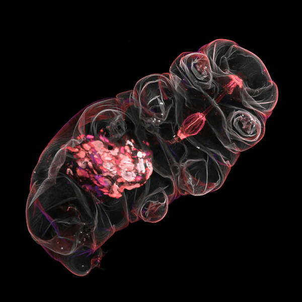
“Tardigrade.”
Image Of Distinction: Jonathan Muyal, Paris, France

“Iridescent rutile (mineral) needles in a Burmese ruby.”
Image Of Distinction: Dr. Michael Weber, Berlin Institute Of Health At Charité, Department Of Human Genetics, Berlin, Germany
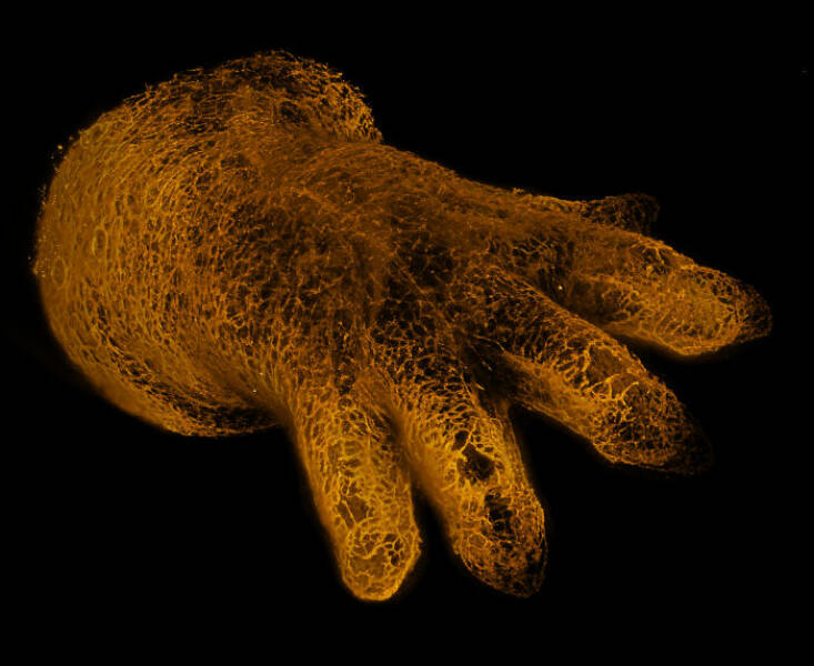
“Blood vessels in the limb of an embryonic mouse.”
Image Of Distinction: Dr. Stephen De Lisle, Karlstad University, Department Of Environmental And Life Sciences, Karlstad, Värmland, Sweden
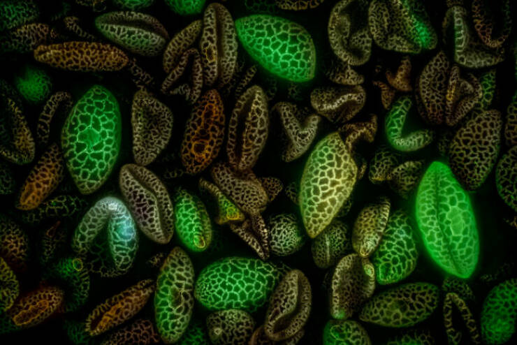
“Lily flower pollen (autofluorescence).”
Image Of Distinction: Walter Ferrari, Walter Ferrari Macro, Rio Cuarto, Cordoba, Argentina
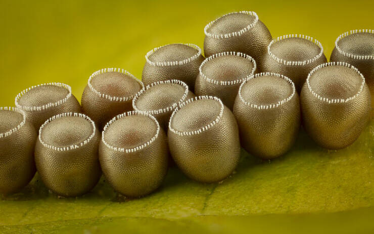
“True bug (Hemipteran) eggs on a leaf.”
16th Place: Rogelio Moreno, Panama, Panama
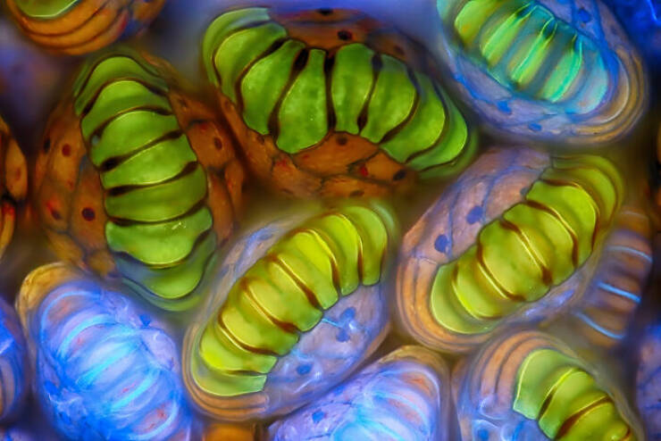
“Spore sacs (sporangia) of a fern.”
Honorable Mention: Dr. Bruno Cisterna And Dr. Eric Vitriol, Medical College Of Georgia At Augusta University, Department Of Neuroscience & Regenerative Medicine, Augusta, Georgia, USA
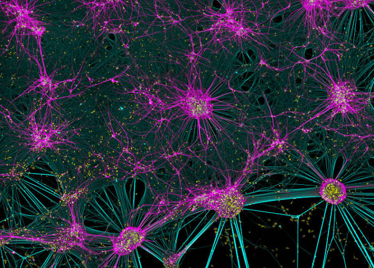
“Human neurons reprogrammed from skin cells.”
Image Of Distinction: Jianguo Mao, Shanghai, Shanghai, China
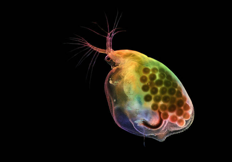
“Pregnant water flea (Daphnia).”
Image Of Distinction: Marek Miś, Marek Miś Photography, Suwalki, Podlaskie, Poland

“Air bubbles in melted polyvinyl alcohol.”
Image Of Distinction: Dr. Mette Handberg-Thorsager, Alexandre Alié And Lisa Maria Ulbrich, Georg-August-University Göttingen, Department Of Multiscale Biology, Göttingen, Niedersachsen, Germany
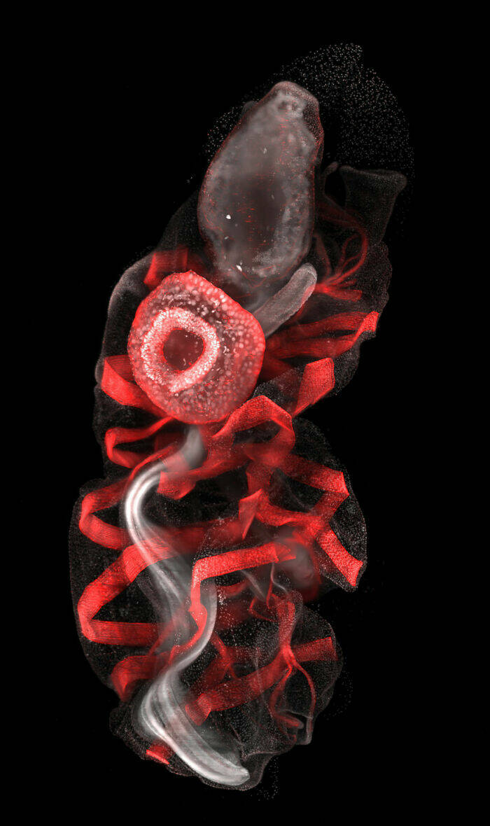
“Oozoid of a sea squirt (Thalia democratica).”
15th Place: Zhang You, Kunming, Yunnan, China
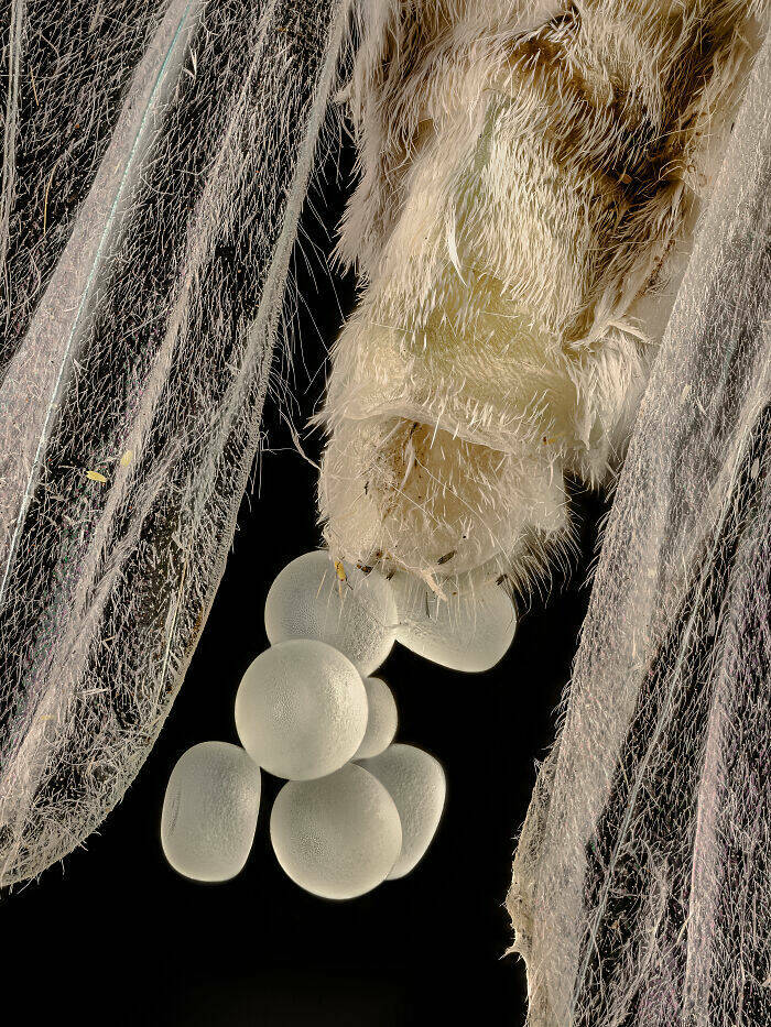
“Geometer moth (Geometridae) laying eggs.”
Image Of Distinction: Bernard Allard, Club Français De Microscopie, Sucy-En-Bry, France
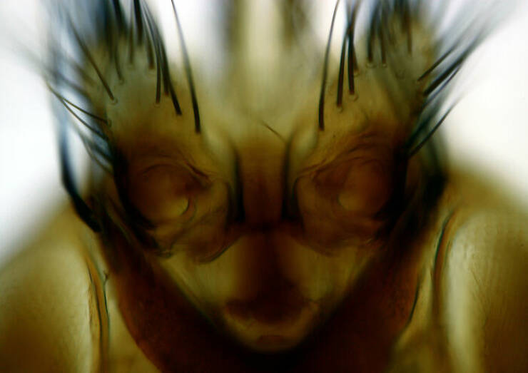
“Parasitic fly (Crataerina hirundinis).”
Image Of Distinction: Joe Mckellar, Cnrs, The Institue Of Molecular Genetics Of Montpellier (Igmm), Montpellier, Hérault, France
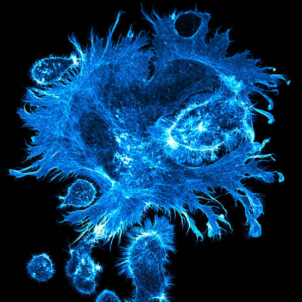
“Giant human hepatic cancer cell surrounded by smaller cells.”
Image Of Distinction: Dr. Noah Bressman, Salisbury University, Department Of Biology, Salisbury, Maryland, USA
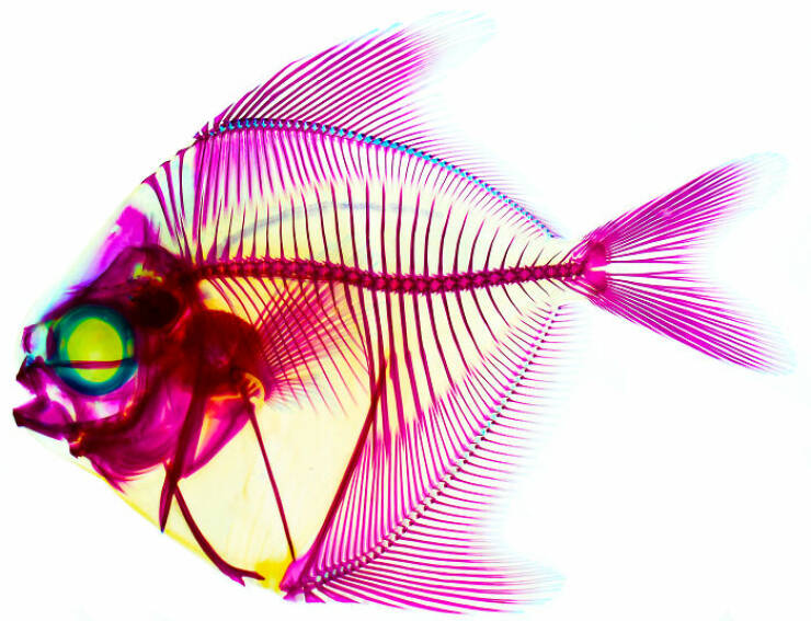
“Histologically-stained harvestfish/star butterfish (Peprilus paru).”
Image Of Distinction: Solvin Zankl, Kiel, Schleswig-Holstein , Germany
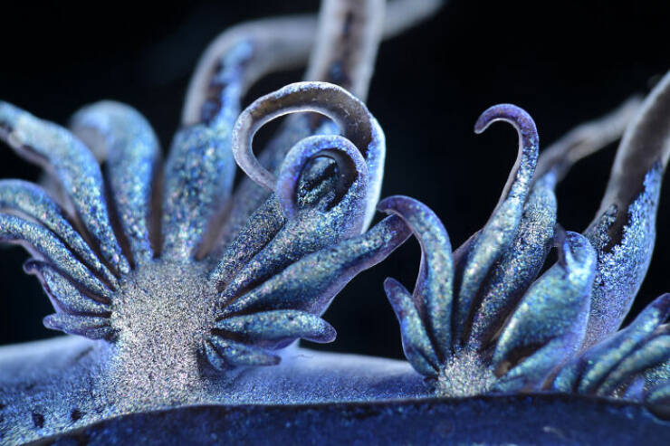
“A floating sea slug (Glaucus atlanticus, also known as the blue sea dragon).”
9th Place: Wim Van Egmond, Micropolitan Museum, Berkel En Rodenrijs, Zuid Holland, Netherlands
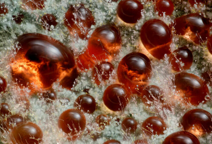
“A fungus (Talaromyces purpureogenus) known for its red, diffused pigment.”
19th Place: Eduardo Carrasco, Cuenca, Azuay, Ecuador
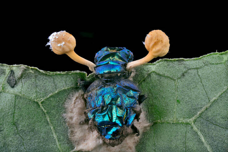
“Parasitic fungus (Cordycipitaceae) on a fly (Calliphoridae).”
Image Of Distinction: Frederic Labaune, Education Nationale, Auxonne, Burgundy, France
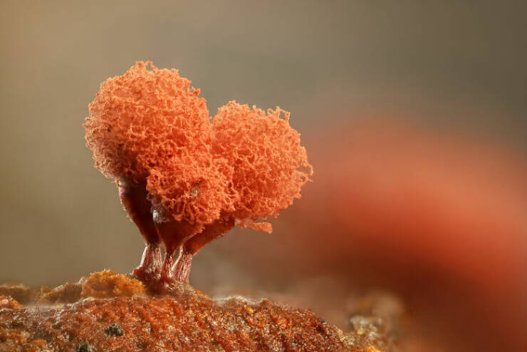
“Slime mold (Arcyria denudata).”
Image Of Distinction: Dr. Laurent Formery, University Of California Berkeley, Department Of Molecular And Cell Biology, Pacific Grove, California, USA
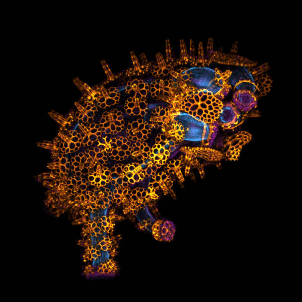
“Skeleton of a juvenile sea cucumber.”
Image Of Distinction: Daniel Han, Diatoms Australia (Macro Cosmos Imaging), Sydney, New South Wales, Australia
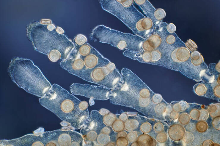
“Diatoms (Arachnoidiscus sp.) on coralline algae.”
Image Of Distinction: Doong Yien, Xmato Works, Beijing, China
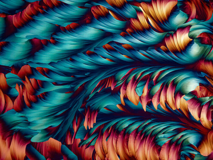
“Crystallization of a mixed solution of alanine and glutamine under polarized light.”
Image Of Distinction: Heiti Paves, Tallinn, Harju, Estonia
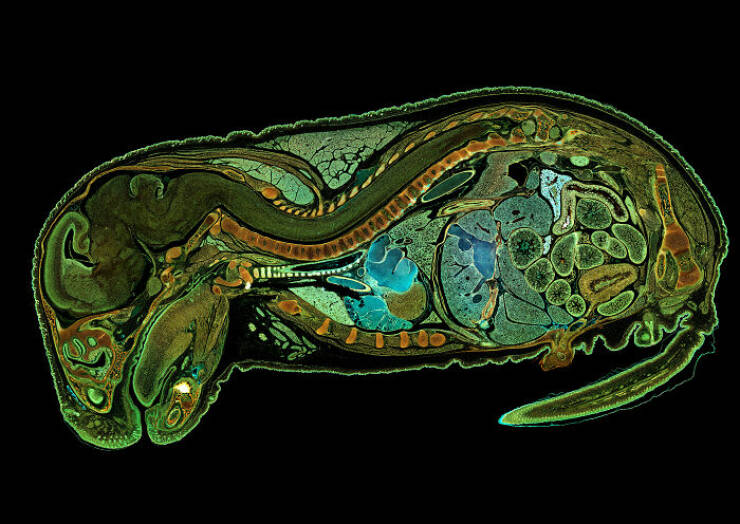
“Mouse embryo, sagittal section.”
Image Of Distinction: Dr. Julien Resseguier, University Of Oslo, Department Of Biosciences / Fyscell, Oslo, Viken, Norway
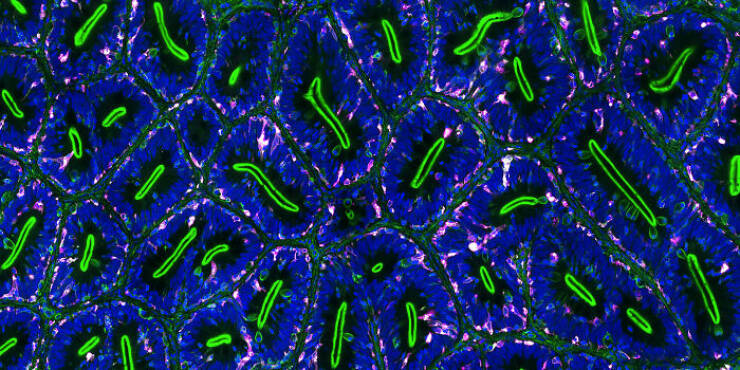
“Immune cells (magenta) protecting the different tissue compartments of the zebrafish intestines.”
Image Of Distinction: Robert Schmittling, Hillsborough, North Carolina, USA
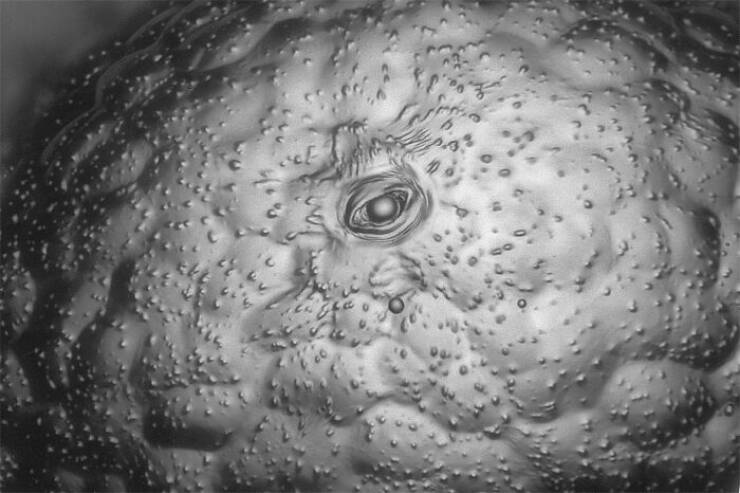
“Eye of potato (stomate).”
Image Of Distinction: Stephanie Huang, Victoria University Of Wellington, School Of Biological Sciences; School Of Psychology, Wellington, New Zealand
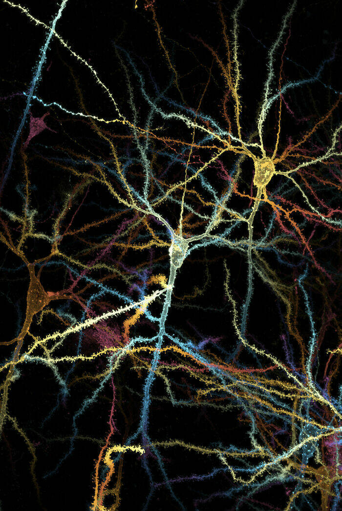
“Pyramidal neurons from the ventral orbital cortex (prefrontal cortex) from an adult rat brain.”
Honorable Mention: Dr. Frédéric Fercoq And Jean-Gabriel Rothan, Muséum National D’histoire Naturelle, Paris, France

“Larvae of a filarial parasite (nematode).”
Honorable Mention: Rebecca Lee, Yale University, Department Of Genetrics, New Haven, Connecticut, USA
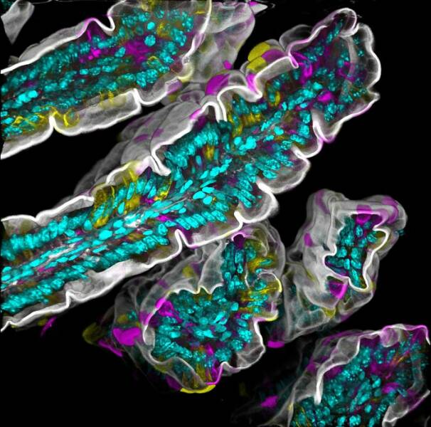
“Villi in the mouse small intestine.”
Image Of Distinction: Bilal Akhtar, Institute Of Molecular Biology, Neuroscience-Rna Biology, Mainz, Rheinland-Pfalz, Germany
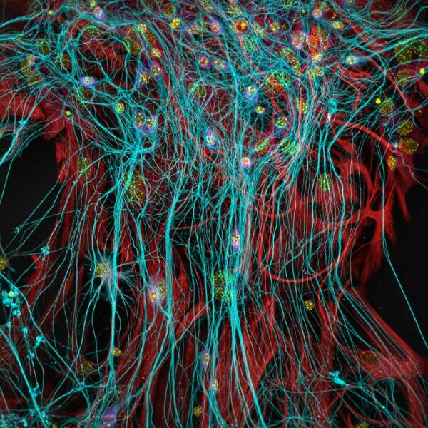
“14-day-old mouse neuronal co-culture with astrocytes.”
Image Of Distinction: Hannah Somers, Mdi Biological Laboratory, Light Microscopy Facility, Bar Harbor, Maine, USA
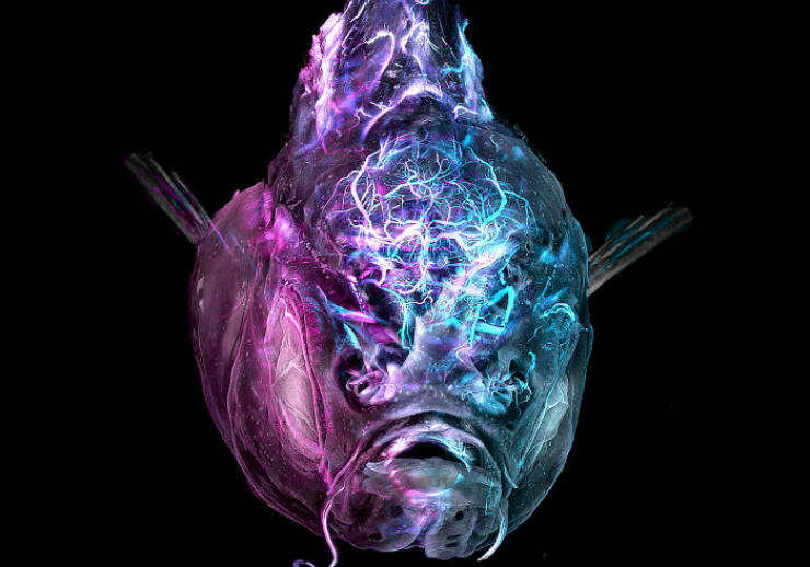
“An adult zebrafish showing blood vessels in the brain.”
Image Of Distinction: Shambhavi Dwivedi And Dr. Friedemann Kiefer, University Of Münster, Faculty Of Biology, Muenster, North-Rhine Westphalia, Germany
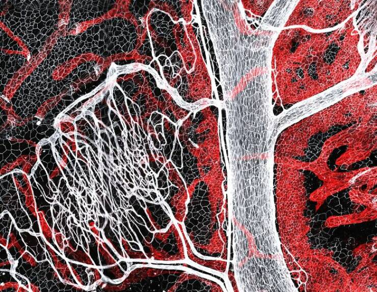
“Mouse lymphatic network (red) flanking blood vessels (white).”
4th Place: Dr. James Hayes, Vanderbilt University, Department Of Cell And Developmental Biology, Nashville, Tennessee, USA
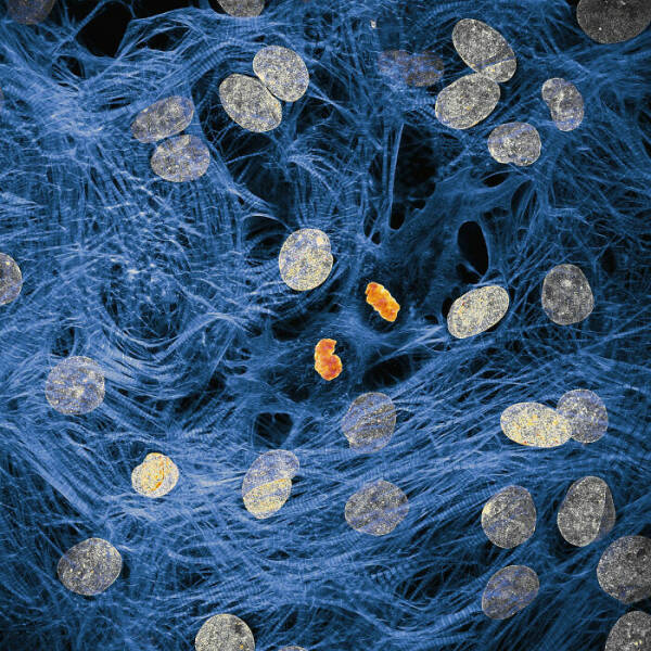
“Heart muscle cells with chromosomes condensed following cell division.”
10th Place: Dr. Dylan Burnette And Dr. James Hayes, Vanderbilt University School Of Medicine, Department Of Cell And Developmental Biology, Nashville, Tennessee, USA
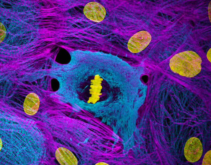
“Heart muscle cells (iPSC-derived) showing condensed chromosomes in metaphase.”
Honorable Mention: Mishal Abdulaziz Alryhan, Fiap, Al-Ahsa, Saudi Arabia
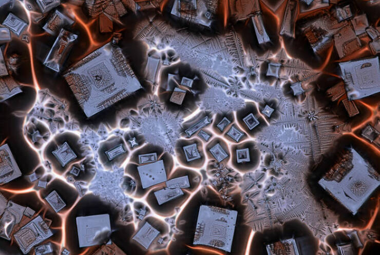
“Crystallized soy sauce fusion with alum.”