 Barnorama All Fun In The Barn
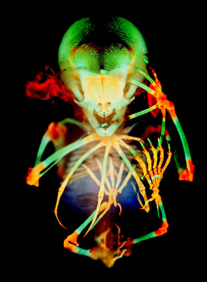
20th place: Dr. Dorit Hockman, Dr. Vanessa Chong-Morrison. Skeleton preparation of a short-tailed fruit bat embryo (Carollia perspicillata).
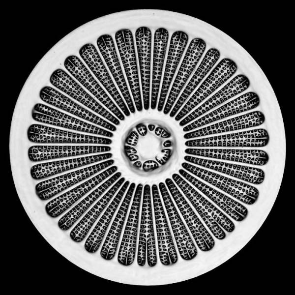
19th place: Dr. Jan Michels. Silica cell wall of the marine diatom Arachnoidiscus sp.
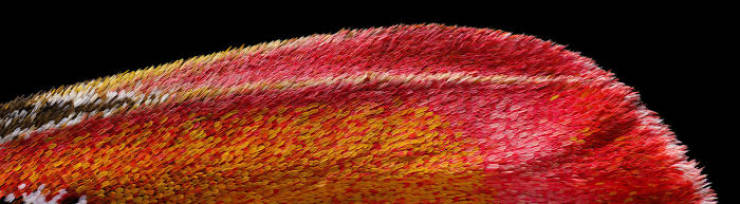
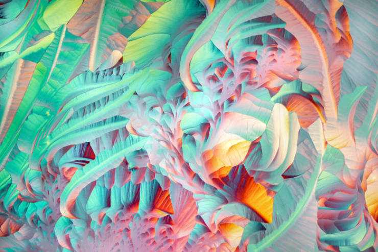
18th place: Chris Perani. Atlas moth wing.
17th place: Anne Algar. Ventral view of an immature water boatman.
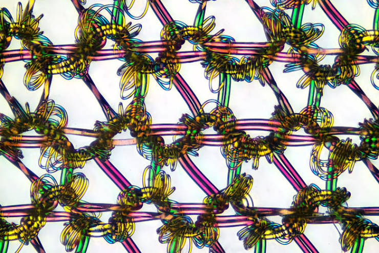
16th place: Alexander Klepnev. Nylon stockings.
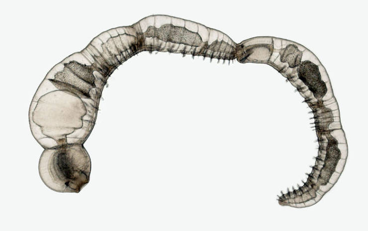
15th place: Dr. Eduardo Zattara, Dr. Alexa Bely. Chain of daughter individuals from the asexually reproducing annelid species Chaetogaster diaphanus.
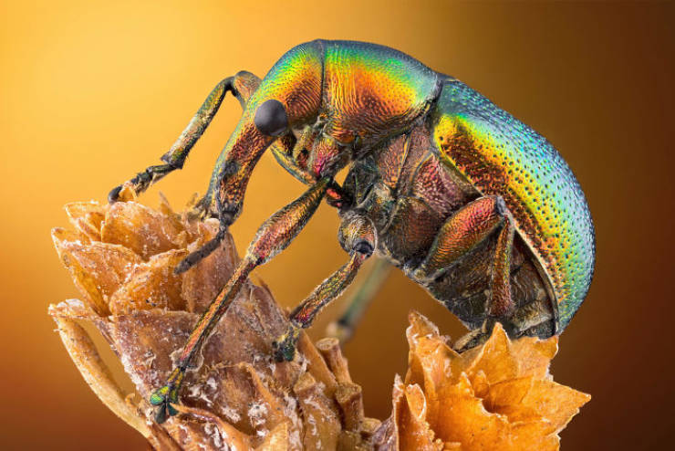
14th place: Özgür Kerem Bulur. Leaf roller weevil (Byctiscus betulae) lateral view.
13th place: Justin Zoll. Crystals formed after heating an ethanol and water solution containing L-glutamine and beta-alanine.
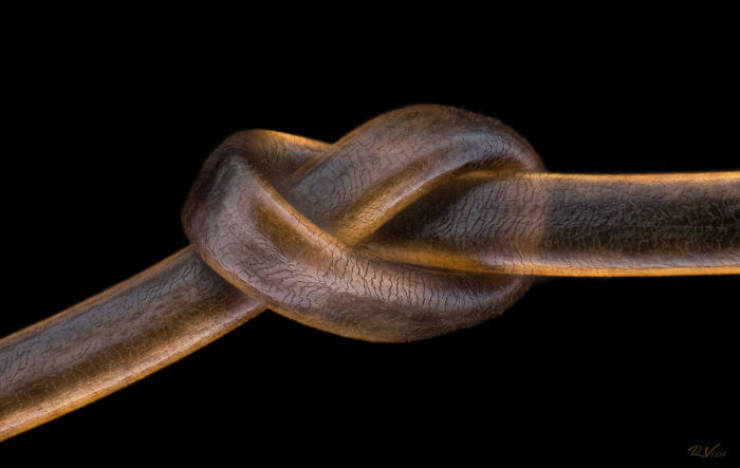
12th place: Robert Vierthaler. Human hair.
11th place: Dr. Tagide deCarvalho. Red algae.
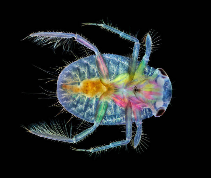
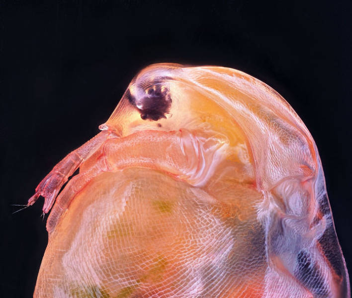
10th place: Ahmad Fauzan. Daphnia magna (Phyllopoda).
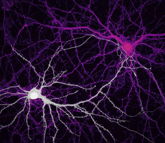
9th place: Jason Kirk, Quynh Nguyen. Connections between hippocampal neurons (brain cells).
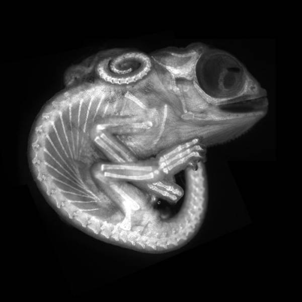
8th place: Dr. Allan Carrillo-Baltodano, David Salamanca. Chameleon embryo (autofluorescence).
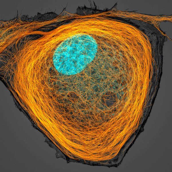
7th place: Jason Kirk. Microtubules (orange) inside a cell. Nucleus is shown in cyan.
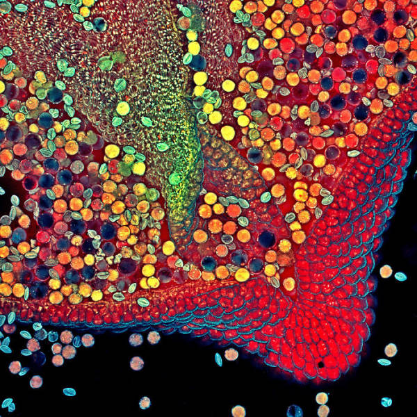
6th place: Dr. Robert Markus, Zsuzsa Markus. Hebe plant anther with pollen.
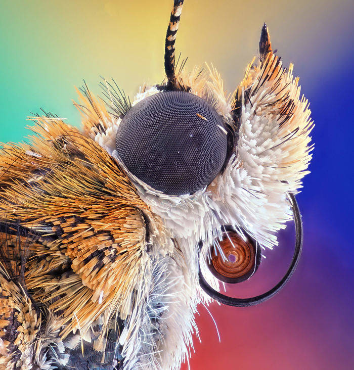
5th place: Ahmad Fauzan. Bogong moth.
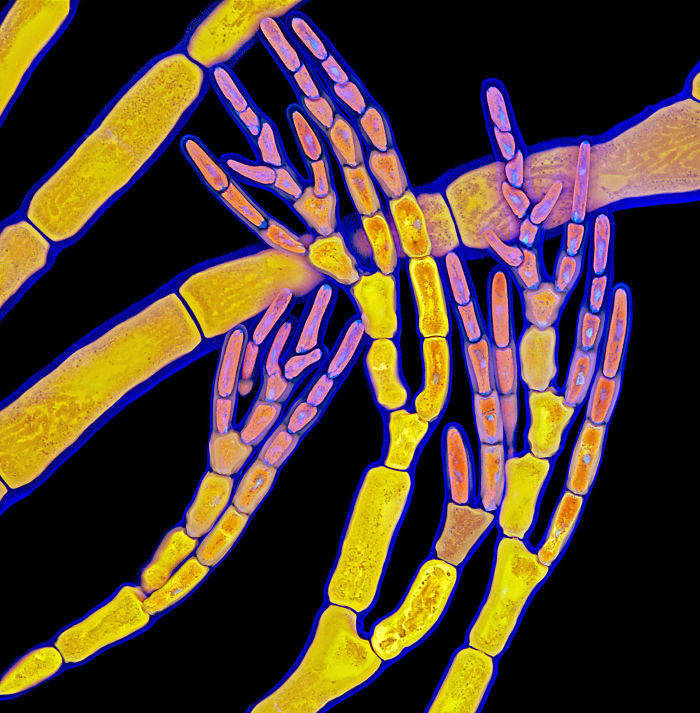
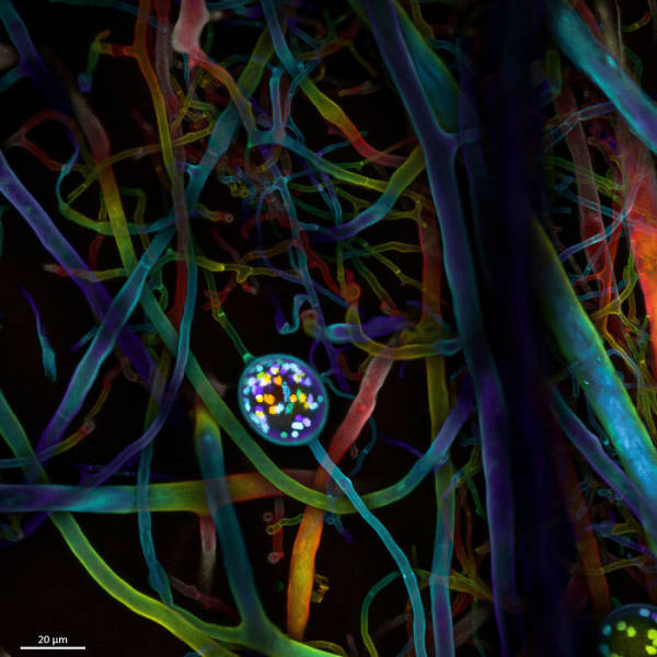
4th place: Dr. Vasileios Kokkoris, Dr. Franck Stefani, Dr. Nicolas Corradi. Multi-nucleate spores and hyphae of a soil fungus (arbuscular mycorrhizal fungus).
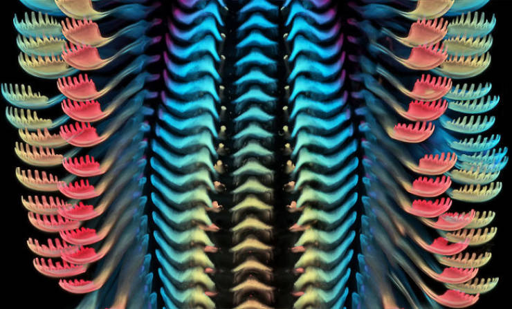
3rd place: Dr. Igor Siwanowicz. Tongue (radula) of a freshwater snail.
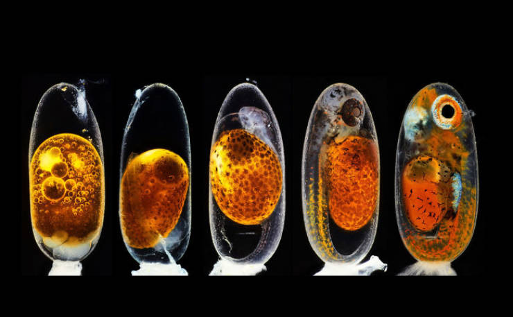
2nd place: Daniel Knop. Embryonic development of a clownfish (Amphiprion percula) on days 1, 3 (morning and evening), 5, and 9.
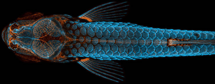
1st Place: Daniel Castranova, Dr Brant Weinstein & Bakary Samasa. Dorsal view of bones and scales (blue) and lymphatic vessels (orange) in a juvenile zebrafish.